Gallery
Making a measurement and making an image are the same gesture performed twice. These images came out of experiments and went on to be recognised in image competitions or appear on a journal cover.
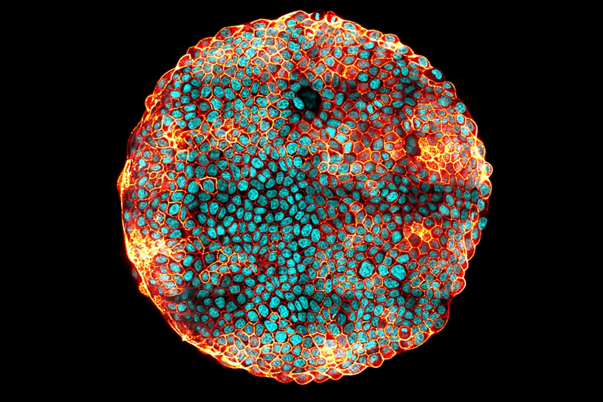
An island of MDCK epithelial cells, several thousand strong, confined to a circular patch. Nuclei appear in cyan, cell-cell junctions in red and orange. At the rim, where the tissue meets free space, cells crowd and their junctions brighten (the signature of the mechanical tension the edge carries). Exhibited at the Cité des sciences et de l'industrie, Paris.
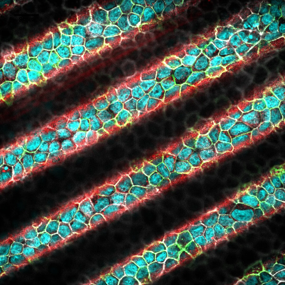
An MDCK epithelium spread over a substrate corrugated into regular waves. A single confocal plane cuts through the crests, so the layer appears as diagonal stripes: nuclei in cyan, cell-cell junctions in green and white, and the crest lines picked out in red. How the thickness of the tissue varies along these waves is what the paper set out to measure, and the image made the cover of the issue.
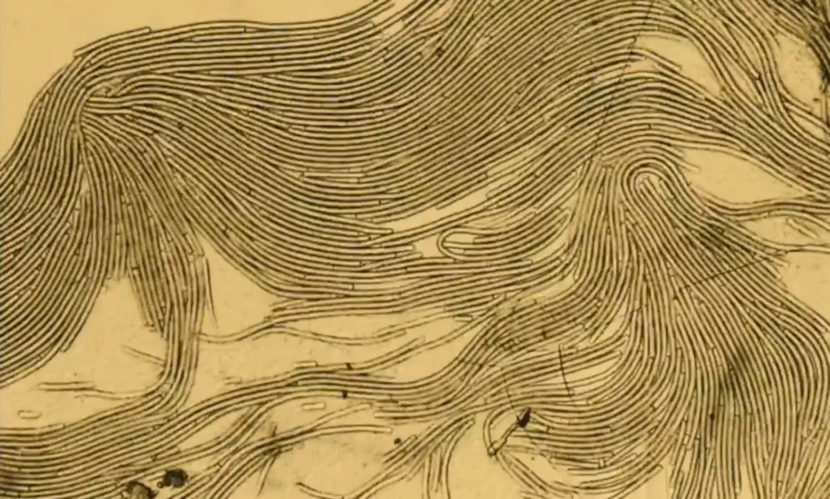
Oscillatoria princeps, a filamentous cyanobacterium that glides across surfaces. Left to their own devices, the filaments align with their neighbours and organise into swirls and defects (an experimental realisation of an active nematic liquid crystal, made of living matter).


