Stress sensors and rheology measurements in living tissues
Understanding how mechanical forces shape tissues during embryonic development requires measuring their physical properties in vivo. Conventional macroscopic rheology cannot do this: it is destructive, and it does not reach cellular scales.
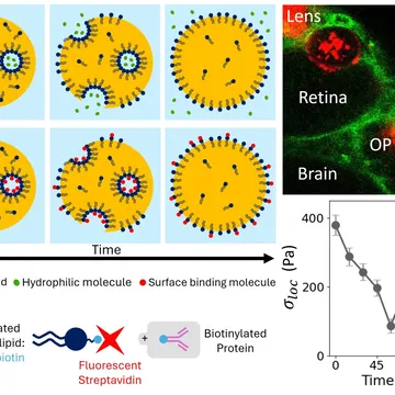
We inject ferrofluid droplets directly into the extracellular matrix, or between cells of the embryo. Applying controlled oscillating magnetic fields deforms these droplets, and their deformation gives a local, non-destructive read-out of the mechanical response (viscoelasticity, plasticity) of the surrounding matrix.
The approach maps the spatiotemporal dynamics of shear forces and mechanical stresses during morphogenesis.

Collaborations
- Experimental biophysics : L.-L. Pontani, E. Wandersman & A. Prevost (LJP)
- Cell & developmental biology : M.-A. Breau & C. Curantz (Dev2A)
- Theory & modelling : J. O'Byrne (LJP)
Related publications
-
2026
Design and optimization of in situ self-functionalizing stress sensors The European Physical Journal E 49, 12 (2026)*Equal contributions DOI: 10.1140/epje/s10189-025-00550-y